Growth and nourishment of bone. Unlike cartilage, bone has a very good blood supply. Bone is riddled with blood capillaries. The central cavity contains blood vessels and is a storage for bone marrow.
What is the blood and nerve supply of bones?
Haversian canals typically run parallel to the surface and along the long axis of the bone and generally contain one or two capillaries and nerve fibers. Volkmann’s canals are channels that assist with blood and nerve supply from the periosteum to the Haversian canal.
Why are there nerves in the bone?
Essentially, bone nerves have been implicated in two different roles: as regulators of bony mechanical forces and as a source of trophic factors essential for structure and bone function. According to Wolff’s law, different grades of physical activity are converted into changes in bone mass.
Are there nerves in bones?
Bone tissue contains a dense network of sensory and sympathetic nerve fibers, which appears to play important roles in bone modeling, remodeling, metabolism, and adaptation (84).How are the bones nourished?
Blood and Nerve Supply The osteocytes in spongy bone are nourished by blood vessels of the periosteum that penetrate spongy bone and blood that circulates in the marrow cavities. As the blood passes through the marrow cavities, it is collected by veins, which then pass out of the bone through the foramina.
What is the tubercle of a bone?
A tubercle is a small rounded point of a bone. It also refers to a nodule attached to bone, mucous membrane (moist layer lining parts of the body), or skin.
How do bones get nourishment?
The blood supply to bone is delivered to the endosteal cavity by nutrient arteries, then flows through marrow sinusoids before exiting via numerous small vessels that ramify through the cortex.
What is Volkmann's canal?
[ fōlk′mənz, -mänz′ ] n. Any of the various canals in bone that transmit blood vessels from the periosteum into the bone.What canal connects the periosteum to the haversian Canal?
Volkmann’s canals, also known as perforating holes or channels, are anatomic arrangements in cortical bones. Volkmann’s canals are inside osteons. They interconnect the haversian canals with each other and the periosteum.
How are bones innervated?Bone tissue is innervated by both myelinated (A beta and A delta fiber) and unmyelinated (C fiber) sensory neurons. In combination, they can provide an initial burst of pain, initiated by the faster myelinated fibers, followed by a slower and longer-lasting dull pain initiated by unmyelinated fibers.
Article first time published onIs cartilage innervated?
Cartilage has no nerve innervation, and hence there is no sensation when it is injured or damaged. When there is calcification of cartilage, the chondrocytes die. This is followed by the replacement of cartilage with bone-like tissue. Unlike bone, cartilage does not have calcium in the matrix.
Are teeth bones?
Even though teeth and bones seem very similar, they are actually different. Teeth are not bones. Yes, both are white in color and they do indeed store calcium, but that’s where their similarities end.
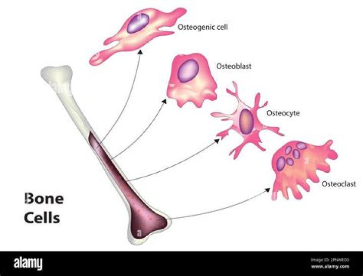
What are bone producing cells?
Osteoblasts are bone-forming cells, osteocytes are mature bone cells and osteoclasts break down and reabsorb bone.
What supplies blood to the bone?
The blood supply to bone is delivered to the endosteal cavity by nutrient arteries, then flows through marrow sinusoids before exiting via numerous small vessels that ramify through the cortex.
Which blood vessel supplies the diaphysis of a long bone?
The diaphysis and metaphysis are nourished primarily by the nutrient artery, which passes through the cortex into the medullary cavity and then ramifies outward through haversian and Volkmann canals to supply the cortex.
What is the purpose of the diaphysis of a bone?
The diaphysis contains the medulla of the bone, which houses bone marrow. The marrow is the primary tissue responsible for the production of erythrocytes, leukocytes, and platelets. The epiphysis is the terminal end of the long bone that is typically responsible for articulation.
What are the cellular components of bone tissue?
Bone is composed of four different cell types; osteoblasts, osteocytes, osteoclasts and bone lining cells. Osteoblasts, bone lining cells and osteoclasts are present on bone surfaces and are derived from local mesenchymal cells called progenitor cells.
What type of bone makes up the majority of the diaphysis of long bones like the humerus?
What type of bone makes up the majority of the diaphysis of long bones like the humerus? Describe the layers of bone tissue found here. Most of the bone tissue of the humerus is compact bone. Compact bone is on the outside and spongy (cancellous) bone is on the inside.
How do nutrients enter and metabolites leave the cartilage?
The cells of cartilage, called chondrocytes, are isolated in small lacunae within the matrix. Although cartilage is avascular, gaseous metabolites and nutrients can diffuse through the aqueous phase of the gel-like matrix to reach the cells.
What part of the bone structure contributes to nourishing the underlying bone?
The Periosteum, a fibrous membrane, covers the outside of bone. This membrane is rich with capillaries, which are responsible for nourishing bone.
Where does bone get its strength from?
Made mostly of collagen, bone is living, growing tissue. Collagen is a protein that provides a soft framework, and calcium phosphate is a mineral that adds strength and hardens the framework. This combination of collagen and calcium makes bone strong and flexible enough to withstand stress.
What are bone markings and why are they important?
Bone markings are projections and depressions found on bones, which help us to identify the location of other body structures, such as muscles. Their importance comes when we try to describe the shape of the bone or to understand how the muscles, ligaments and other structures affect this bone and vice versa.
How does a tubercle form?
Tubercles are nodules that contain caseous necrosis, which form in the lungs as a result of an infection with Mycobacterium tuberculosis in the patients with tuberculosis. Granulomas form in the infected tissue and undergo necrosis in the centre. Tubercles are also known as tuberculous nodules, or tuberculomas.
What are bone surface markings?
There are three general classes of bone markings: (1) articulations, (2) projections, and (3) holes. As the name implies, an articulation is where two bone surfaces come together (articulus = “joint”). … A hole is an opening or groove in the bone that allows blood vessels and nerves to enter the bone.
Is cartilage vascular and innervated?
Cartilage is a flexible connective tissue that differs from bone in several ways; it is avascular and its microarchitecture is less organized than bone. Cartilage is not innervated and therefore relies on diffusion to obtain nutrients.
When does the epiphyseal plate replace bone?
When bone growth is complete, the epiphyseal cartilage is replaced with bone, which joins it to the diaphysis. Fractures of the epiphyseal plates in children can lead to slow bone growth or limb shortening. The coordinated activity of these bone cells allows bone to grow, repair itself, and change shape.
Where do osteocytes live?
In mature bones, osteocytes and their processes reside inside spaces called lacunae (Latin for a pit) and canaliculi, respectively. Osteocytes are simply osteoblasts trapped in the matrix that they secrete.
Do bones have layers?
Bones are composed of two layers: a tough outer layer and a spongy inner layer. The outer layer, known as cortical or compact bone, is strong and dense. The inner layer, known as trabecular or cancellous bone, features a light network of connective tissue.
How do osteoblasts form new bone?
4.2. Osteoblasts are the bone cells derived from osteochondral progenitor cells that form the bone through a process called ossification. Osteoblasts result in the formation of new layers of bone by producing a matrix that covers the older bone surface.
Which structure is joined by Volkmann's canal?
function in bone vascular system …of the cortex, are called Volkmann canals; Volkmann canals connect adjacent osteons and also connect the blood vessels of the Haversian canals with the periosteum, the tissue covering the bone’s outer surface.
What type of connective tissue provides nourishment to the bone?
Subchondral tissue. The tough, thin outer membrane covering the bones is called the periosteum. Under the hard outer shell of the periosteum are tunnels and canals. Through these, blood and lymphatic vessels carry nourishment for the bone. Muscles, ligaments, and tendons may attach to the periosteum.